What is Cruciate Ligament Disease?
 Cruciate ligament disease is a common condition of dogs. The cranial cruciate ligament (CCL) is an important ligament within the knee (stifle) responsible for keeping the shin bone (tibia) from sliding forwards in front of the thigh bone (femur).
Cruciate injuries in dogs are almost always the result of progressive weakening and degeneration of the ligament, which is different to people where it is usually the result of an acute injury. This difference in pathology accounts for the very different approaches to treatment options between people and dogs. It is also the reason that a large proportion of dogs who suffer CCL disease in one stifle will develop the same condition in the other leg at some point in their life. The instability within the stifle can also result in damage to another structure within the joint, the meniscus, which can become torn as the tibia and femur move in relation to each other.
What are the signs of cruciate ligament disease?
Dogs suffering from CCL disease will often show lameness in their back legs. This lameness can either be sudden in onset, or a gradual stiffness when getting up after lying down, particularly after exercise. On occasion a ‘clicking’ noise can be heard from the stifle joint.
How is CCL disease diagnosed?
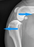
Orthopaedic examination is often enough to give a a provisional suspicion of CCL disease. The stifle joint will often feel thickened and swollen, there is often pain on manipulation of the stifle and in some cases muscle wastage over the affected leg can be felt. X-rays are very useful in assessing the joints, and x-rays of both back legs and hips will be taken to check for other causes for lameness. Sometimes a sample of joint fluid from the knee is taken to help with diagnosis and rule out infection or other inflammatory conditions.
How is CCL disease treated?
Whilst most dogs will benefit from surgery, some dogs with very mild signs can sometimes be managed conservatively with exercise management, physiotherapy, hydrotherapy and pain management.
There are several surgical options available to treat CCL disease. The most commonly performed surgery with us is the TPLO procedure (tibial plateau levelling osteotomy).
TPLO surgery aims to change the angle on the top of tibia to reduce the movement between the tibia and the femur. This is performed by performing a curved cut at the top of the tibia, rotating this portion of bone, and then stabilising it in a new position with a plate and screws.
During surgery we always looking inside the joint. This allows us both to confirm that the cruciate ligament has been damaged as we expect, and also allows us to examine the cartilage pads (menisci) within the joint. There are two of these menisci within the stifle joint, and one is particularly prone to injury following CCL disease. Any torn portion of meniscus will be removed during surgery, and this often makes patients feel a lot more comfortable.
What happens after surgery?
Patients will be hospitalised overnight following to continue with their pain relief and ensure a continued smooth recovery from their anaesthetic. An epidural anaesthetic is given before surgery, and this can prevent them from walking for 8-10 hours after administration. Remaining in hospital allows us to care for them until they are able to safely walk.
Exercise must be restricted post-operatively. Cage rest is strongly recommended. Short walks out to the toilet on a lead are all that is permitted during the first week. A post-operative check up will be scheduled with one of our surgeons 7-10 days after surgery, and increases in controlled lead exercise will be discussed at this point.
Complete return to normal activity will usually take 12-16 weeks.
Overall TPLO is a very successful surgery with low risks of serious complications. Owner satisfaction is generally high and over 75% of dogs have been found to return to full function. It should be noted that these joints are not normal, and we do expect to see an ongoing progression of degenerative joint disease, but by improving joint stability we expect this to occur at a slower rate than without surgery. Complications are uncommon, but can include:
- Haemorrhage; this can occur due to the close proximity of blood vessels to the surgical site, and is addressed during surgery
- Infection; buster collars will be provided to prevent patients from licking at the surgical site, as this is the most common cause for surgical site infection. Severe infections may require the plate and screws to be removed once the tibia has healed
- Fracture; patients must be strictly rested following surgery to help reduce this ris as much as possible